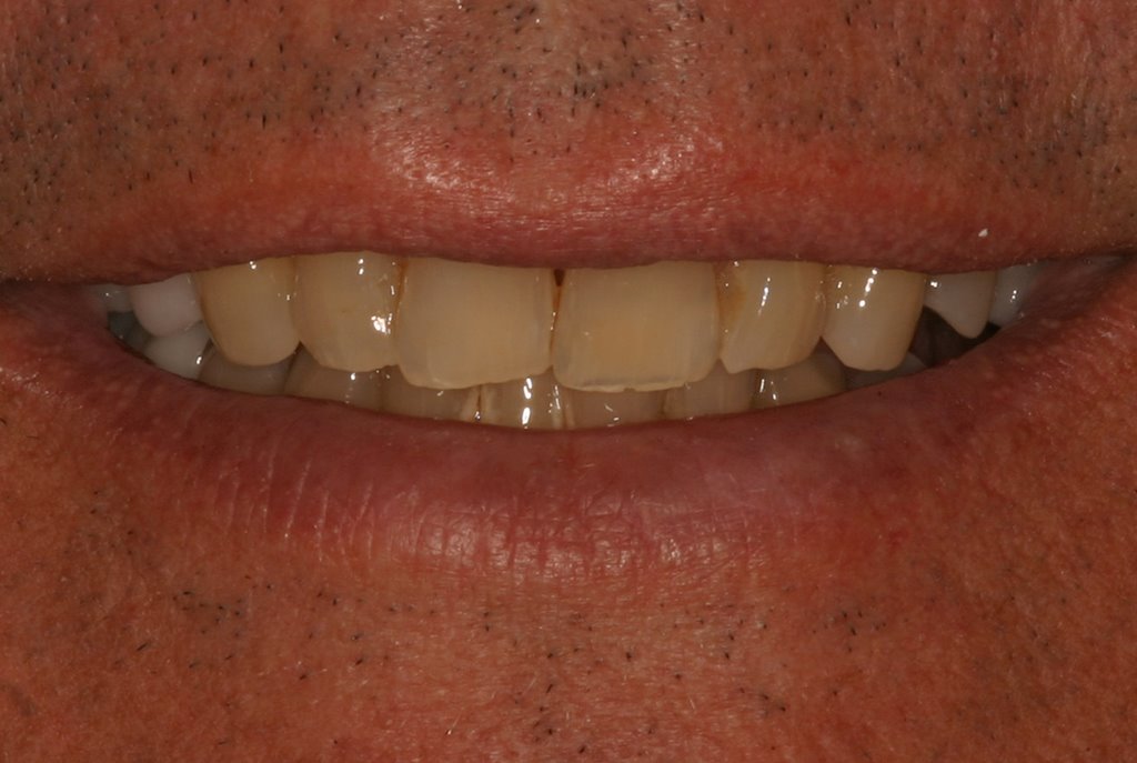
A Patient walks in with the situation below. Five years ago, this would have been a multi-dimensional treatment plan, taking weeks for the tissue to heal after surgery. Luckily for us and the patient, he walked in today at 10:30 am and was out the door at noon, able to eat dinner tonight.
 |
1) PreOp Vertical Fracture
|
After numbing the patient, we removed the fractured portion to find that the clean vertical fracture extended ~3mm below the gingival margin (see picture 2 and 3). Using a Diode Laser, we removed gingival tissue to 1 mm below the planned crown margin (also picture 2 and 3).
 |
2) Occlusal View once the fractured portion was removed
 |
3) Lingual View once the frctured portion was removed
Next we prepped the tooth for a Porcelain Crown, leaving the crown margin above the extent of the fracture. The root surface below this margin was smooth, allowing for long term stability of the periodontal tissues (picture 4 below).
 |
| 4) Prepped tooth to a position where biologic stability will be achieved |
|
|
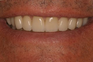
No impressions, just CEREC scans of the upper arch, lower arch, and bite of the patient. Designed the crown, milled the crown, and tried the Porcelain Crown in (pictures 5-8) . No adjustments, polished the Crown, and bonded it in---Final Result below (picture 9). It's awesome to tell a patient that we can fix their major problem in a single visit; they continue on with life as if nothing happened, not realizing that the difference a millimeter makes in one direction or another could have led to a Root Canal, or worse yet, an extraction and implant. One Appointment, 90 minutes, and he'll be ready for dinner with a permanently cemented bonded crown.
 |
| 5, 6, 7) Various Views of the CEREC Crown Designed... |
 |
| 8) Milling preview of the Porcelain Crown |
 |
9) Note: this crown disappears into his smile, yet photographs a bit opaque due to the material used to
functionally stand up to his bite forces |


